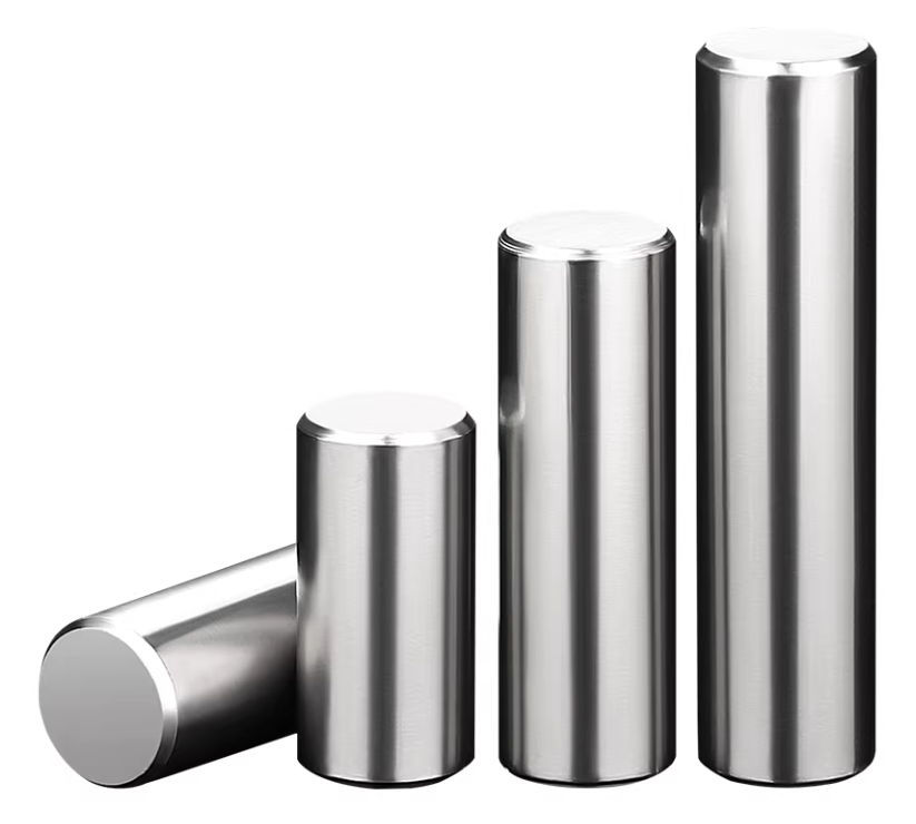
Researchers from Mexico and Costa Rica are working together to advance bone regeneration through bio-3D printing, enabling doctors and surgeons to create patient-specific scaffolds to improve treatment outcomes. For scientists, 3D printing and tissue engineering hold tremendous promise because they offer the ability to precisely fabricate complex geometries. In these experiments, all the classical advantages of 3D printing are also demonstrated, such as affordability, production speed, and optimized performance.
The research team further detailed their findings in the study “Biocompatibility of 3D-Printed Tubular Scaffolds with Nanofibrous Coatings for Bone Applications,” explaining how bone scaffolds can be enhanced by adding composite layers that create surfaces more conducive to cell adhesion and uniform cell seeding. To fabricate these scaffolds, the team employed a unique Air-Jet Spinning (AJS) technique featuring specialized spinning nozzles and a surface for collecting polymer fibers and compressed gas. They also used 3D-printed PLA tubular scaffolds with submicron-fiber surface coatings to evaluate the biological response of human fetal osteoblast cells (hFOB).
This new approach combines a PLA 3D-printed core with an outer layer of nanofibers. The researchers used Cura software for internal geometry slicing and a MakerMex 3D printer to produce the tubular structures. The dual-technology method allowed the team to generate fiber-layer dispersions that formed surfaces with a “uniform thickness distribution,” and the nanofibers integrated well with the 3D-printed scaffold. Adhesion was described as “very strong,” the composites demonstrated increased thermal stability, and the coating endowed the tubular scaffold with properties essential for tissue engineering in bone regeneration.
SEM Micrographs of the 3D-Printed Tubular Scaffold
SEM analysis of the printed tubular scaffold’s 3D surface revealed distinctive morphology and structure, and the surface roughness increased after functionalization with the fiber membrane. Additionally, scaffolds coated with submicron fibers exhibited significantly better hFOB cell adhesion and proliferation compared to uncoated 3D-printed scaffolds. This indicates that the fibers serve as a platform to improve cellular biocompatibility (non-cytotoxicity) and support the attachment and growth of osteoblast cells. The researchers stated that further investigation of the fiber-coated scaffolds is needed to explore their potential role in biomineralization processes and possible future applications in bone tissue engineering or vascularization.

Optical Profilometry Results
Optical profilometry images show the morphology of the 3D-printed tubular scaffolds:
(a) an uncoated scaffold with a smooth surface, and (b) a coated scaffold where surface roughness is significantly increased by the presence of nanofibers.

SEM micrograph of a 3D printed tubular scaffold coated with 7% PLA nanofibers.

*This article originally appeared on China 3D Printing Network. China 3D Printing Network is the original author of this piece.


