

Knowledge Base
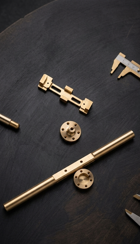
Mechanical Lathe Turning Processing: Complete Analysis from Basics to Advanced Applications | CNC Turning Process Guide
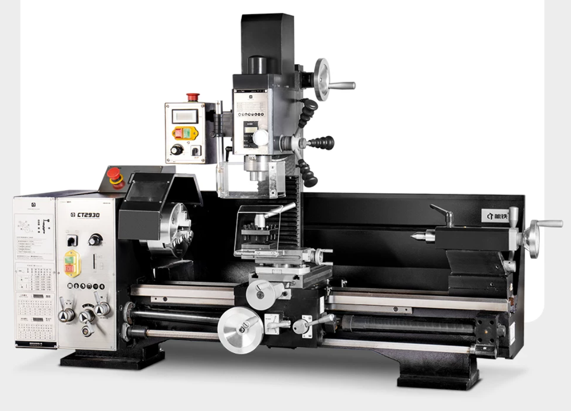
Basic Concepts and Historical Development of Turning Processing What is turning processing? Turning processing is a material removal process that